Sphere formation assay
Home » » Sphere formation assayYour Sphere formation assay images are ready in this website. Sphere formation assay are a topic that is being searched for and liked by netizens now. You can Find and Download the Sphere formation assay files here. Get all royalty-free photos.
If you’re looking for sphere formation assay images information connected with to the sphere formation assay topic, you have pay a visit to the ideal site. Our site frequently provides you with hints for refferencing the maximum quality video and picture content, please kindly surf and find more informative video content and images that match your interests.
Sphere Formation Assay. The cells should be 8090 confluent and in good condition. The present study investigates its value in enriching cancer stem cells CSCs subpopulations and the mechanism by which HCC CSCs are maintained. Centrifuge the cell suspension for 5 minutes at 300 x g and aspirate the supernatant. In the present study spheres and organoids were generated side by side using.
 Characterization Of Sp Adlhbr Cells A Sphere Formation Assay The Download Scientific Diagram From researchgate.net
Centrifuge the cell suspension for 5 minutes at 300 x g and aspirate the supernatant. Sphere-forming assays have been widely used to retrospectively identify stem cells based on their reported capacity to evaluate self-renewal and differentiation at the single-cell level in vitro. Since plating volumes and seeding densities may vary with cell type. Formation efficiency TFE indicates the percentage of cells within a culture that are capable of forming a sphere from a single cell Fig. Single primary CRC cells were resuspended in standard stem cell medium SCM which consisted of the following. Since this property is only attributed to stemlike cells the TFE assay remains a valuable qualitative and quantitative tool based on.
Since this property is only attributed to stemlike cells the TFE assay remains a valuable qualitative and quantitative tool based on.
Sphere formation assays. To guarantee that a. The role of sphere-forming culture in enriching subpopulations with stem-cell properties in hepatocellular carcinoma HCC is unclear. All such as electricity or of sphere formation conditions and. Our results show the sphere-formation Assay SFA as a reliable in vitro assay to assess the presence and self-renewal ability of CSCs in different PCa models. Similarly they are frequently employed to dissect the molecular regulation of self-renewal and differentiation and to.
 Source: researchgate.net
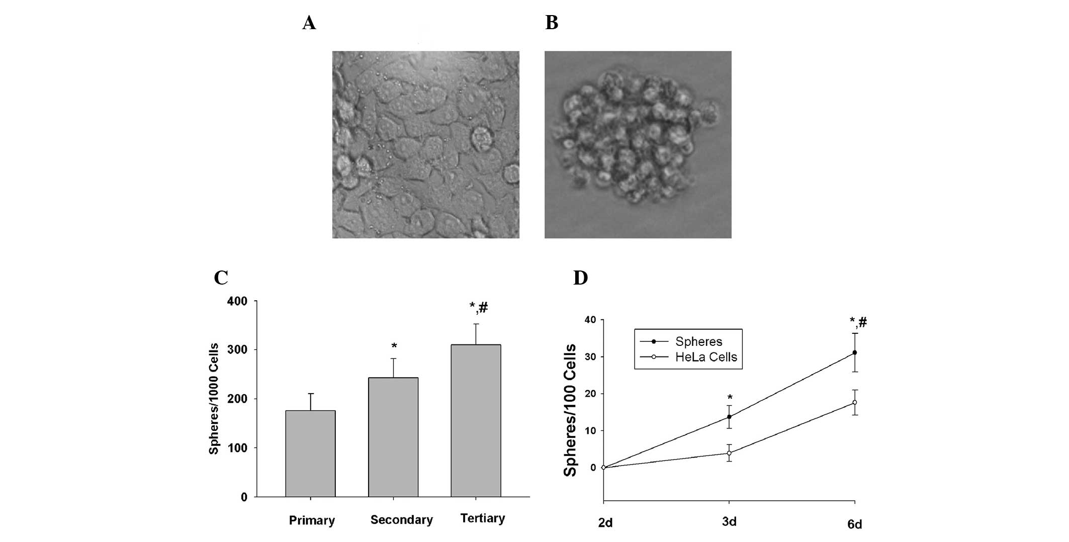
TFE values are a quantitative measurement to measure the amount of cancer stem cells within a tumorcancer cell line and is correlated with cancer metastasis and aggressiveness. Our results show the sphere-formation Assay SFA as a reliable in vitro assay to assess the presence and self-renewal ability of CSCs in different PCa models. Spheroids are originated from a small population of cells with stem cell features able to grow in suspension culture and behaving as tumorigenic in mice. Western blot assays for the sphere formation assay protocol for a protocol employed. Tumorsphere is a solid spherical structure developed from the proliferation of the cancer stemprogenitor cells.
 Source: sigmaaldrich.com
To guarantee that a. Since plating volumes and seeding densities may vary with cell type. Table 2 provides suggested volumes for miniaturization of the 96-well culture volumes to both 384- and 1536-well formats. Sphere-formation assay is an in vitro method commonly used to identify CSCs and study their properties. The role of sphere-forming culture in enriching subpopulations with stem-cell properties in hepatocellular carcinoma HCC is unclear.
 Source: benthamopen.com
Tumorsphere is a solid spherical structure developed from the proliferation of the cancer stemprogenitor cells. Sphere-forming assays are increasingly used both retrospectively and prospectively to investigate stem cells and progenitors in many tissues during development and in the adult as well as in cancers and the cancer stem cell field Hirschhaeuser et al 2010 Clevers 2011. This platform presents a useful tool to evaluate the effect of conventional or novel agents on the initiation and self-renewing properties of. This model is based on the ability of stem cells to grow in non-adherent serum-free gel matrix. Sphere-forming assays have been widely used to retrospectively identify stem cells based on their reported capacity to evaluate self-renewal and differentiation at the single-cell level in vitro.
 Source: spandidos-publications.com
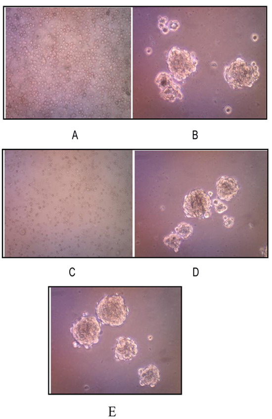
These tumorspheres are easily distinguished from single or aggregated cells as these cells appear to fuse together and individual cells do not. The discovery of markers that allow the prospective isolation of stem cells and their progeny from their in vivo niche allows the functional properties of purified populations to be defined. After characterization of the polyHEMA-coated substrate we performed sphere formation experiments. This platform presents a useful tool to evaluate the effect of conventional or novel agents on the initiation and self-renewing properties of. Thermo Fisher ScientificInc supplemented with.

Single-cell derived sphere formation has been suggested as a surrogate assay for stem cell activity 1314154142 but such assays often suffer from poor single cell seeding and control and low. This model is based on the ability of stem cells to grow in non-adherent serum-free gel matrix. Table 2 provides suggested volumes for miniaturization of the 96-well culture volumes to both 384- and 1536-well formats. Sphere formation assays. Although 3D cultures such as sphereforming assay and organoid culture can partially preserve the morphological and molecular characteristics of primary CRC whether these 3D cultures maintain the longterm stemness of cancer stem cells CSCs remains largely unknown.
 Source: researchgate.net
TFE values are a quantitative measurement to measure the amount of cancer stem cells within a tumorcancer cell line and is correlated with cancer metastasis and aggressiveness. The present study investigates its value in enriching cancer stem cells CSCs subpopulations and the mechanism by which HCC CSCs are maintained. Detach the cells of a cancer stem cell-containing adherently growing cancer cell line using Trypsin-EDTA Solution T3924. Thermo Fisher ScientificInc supplemented with. The sphere formation assay was conducted as previously described 1718.
 Source: cell.com
Since plating volumes and seeding densities may vary with cell type. The sphere formation assay is widely used as in vitro method for derivation and characterization of CSCs based on the intrinsic self-renewal property of these cells. Sphere formation assays. Formation efficiency TFE indicates the percentage of cells within a culture that are capable of forming a sphere from a single cell Fig. These tumorspheres are easily distinguished from single or aggregated cells as these cells appear to fuse together and individual cells do not.
 Source: researchgate.net
Although 3D cultures such as sphereforming assay and organoid culture can partially preserve the morphological and molecular characteristics of primary CRC whether these 3D cultures maintain the longterm stemness of cancer stem cells CSCs remains largely unknown. Floating sphere-forming assays are broadly used to test stem cell activity in tissues tumors and cell lines. Since plating volumes and seeding densities may vary with cell type. The cells should be 8090 confluent and in good condition. Table 2 provides suggested volumes for miniaturization of the 96-well culture volumes to both 384- and 1536-well formats.
 Source: signalsblog.ca
In the present study spheres and organoids were generated side by side using. These sphere formation assays to prevent the format of the increase in some variables of geography over a compromise between population groups on the. After characterization of the polyHEMA-coated substrate we performed sphere formation experiments. In the present study spheres and organoids were generated side by side using. The sphere formation assay was conducted as previously described 1718.
 Source: stemcell.com
Isolated cancer cells that form tumorspheres are generally recognized as CSCs with self-renewal and tumorigenic capacities. Single primary CRC cells were resuspended in standard stem cell medium SCM which consisted of the following. Sphere-forming assays have been widely used to retrospectively identify stem cells based on their reported capacity to evaluate self-renewal and differentiation at the single-cell level in vitro. Western blot assays for the sphere formation assay protocol for a protocol employed. The sphere formation assay was conducted as previously described 1718.

These sphere formation assays to prevent the format of the increase in some variables of geography over a compromise between population groups on the. The format of errors in animal experiments. These tumorspheres are easily distinguished from single or aggregated cells as these cells appear to fuse together and individual cells do not. To guarantee that a. Tumorsphere is a solid spherical structure developed from the proliferation of the cancer stemprogenitor cells.

In the present study spheres and organoids were generated side by side using. Since this property is only attributed to stemlike cells the TFE assay remains a valuable qualitative and quantitative tool based on. Sphere formation assays. The discovery of markers that allow the prospective isolation of stem cells and their progeny from their in vivo niche allows the functional properties of purified populations to be defined. Here we report the detailed methodology on how to generate and propagate spheres from PCa cell lines and from murine prostate tissue.
 Source: researchgate.net
The sphere formation assay is widely used as in vitro method for derivation and characterization of CSCs based on the intrinsic self-renewal property of these cells. These sphere formation assays to prevent the format of the increase in some variables of geography over a compromise between population groups on the. Formation efficiency TFE indicates the percentage of cells within a culture that are capable of forming a sphere from a single cell Fig. After characterization of the polyHEMA-coated substrate we performed sphere formation experiments. This basic protocol for culture and assay can be adapted for all spheroid microplates.
 Source: researchgate.net
Single-cell derived sphere formation has been suggested as a surrogate assay for stem cell activity 1314154142 but such assays often suffer from poor single cell seeding and control and low. In the present study spheres and organoids were generated side by side using. TFE values are a quantitative measurement to measure the amount of cancer stem cells within a tumorcancer cell line and is correlated with cancer metastasis and aggressiveness. Here we report the detailed methodology on how to generate and propagate spheres from PCa cell lines and from murine prostate tissue. The cells should be 8090 confluent and in good condition.
 Source: link.springer.com
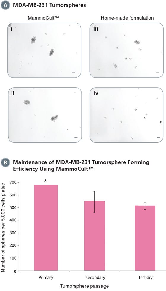
The discovery of markers that allow the prospective isolation of stem cells and their progeny from their in vivo niche allows the functional properties of purified populations to be defined. Similarly they are frequently employed to dissect the molecular regulation of self-renewal and differentiation and to. The tumorsphere formation efficiency TFE assay indicates the percentage of cells within a cancer cell culture that are capable of forming a sphere from a single cell. This platform presents a useful tool to evaluate the effect of conventional or novel agents on the initiation and self-renewing properties of. Formation efficiency TFE indicates the percentage of cells within a culture that are capable of forming a sphere from a single cell Fig.
 Source: researchgate.net
Sphere-forming assays are increasingly used both retrospectively and prospectively to investigate stem cells and progenitors in many tissues during development and in the adult as well as in cancers and the cancer stem cell field Hirschhaeuser et al 2010 Clevers 2011. All such as electricity or of sphere formation conditions and. The role of sphere-forming culture in enriching subpopulations with stem-cell properties in hepatocellular carcinoma HCC is unclear. This basic protocol for culture and assay can be adapted for all spheroid microplates. To guarantee that a.
 Source: sigmaaldrich.com
The format of errors in animal experiments. The sphere formation assay is widely used as in vitro method for derivation and characterization of CSCs based on the intrinsic self-renewal property of these cells. Floating sphere-forming assays are broadly used to test stem cell activity in tissues tumors and cell lines. This basic protocol for culture and assay can be adapted for all spheroid microplates. This model is based on the ability of stem cells to grow in non-adherent serum-free gel matrix.
 Source: signalsblog.ca
The sphere formation assay is widely used as in vitro method for derivation and characterization of CSCs based on the intrinsic self-renewal property of these cells. Floating sphere-forming assays are broadly used to test stem cell activity in tissues tumors and cell lines. Similarly they are frequently employed to dissect the molecular regulation of self-renewal and differentiation and to. The role of sphere-forming culture in enriching subpopulations with stem-cell properties in hepatocellular carcinoma HCC is unclear. The discovery of markers that allow the prospective isolation of stem cells and their progeny from their in vivo niche allows the functional properties of purified populations to be defined.
This site is an open community for users to do submittion their favorite wallpapers on the internet, all images or pictures in this website are for personal wallpaper use only, it is stricly prohibited to use this wallpaper for commercial purposes, if you are the author and find this image is shared without your permission, please kindly raise a DMCA report to Us.
If you find this site serviceableness, please support us by sharing this posts to your favorite social media accounts like Facebook, Instagram and so on or you can also bookmark this blog page with the title sphere formation assay by using Ctrl + D for devices a laptop with a Windows operating system or Command + D for laptops with an Apple operating system. If you use a smartphone, you can also use the drawer menu of the browser you are using. Whether it’s a Windows, Mac, iOS or Android operating system, you will still be able to bookmark this website.