Immunofluorescence staining protocol
Home » » Immunofluorescence staining protocolYour Immunofluorescence staining protocol images are available in this site. Immunofluorescence staining protocol are a topic that is being searched for and liked by netizens today. You can Get the Immunofluorescence staining protocol files here. Find and Download all free photos and vectors.
If you’re looking for immunofluorescence staining protocol images information related to the immunofluorescence staining protocol keyword, you have come to the ideal site. Our site always gives you suggestions for seeking the maximum quality video and picture content, please kindly surf and find more enlightening video content and images that match your interests.
Immunofluorescence Staining Protocol. Preparation of Slides. 10 minutes with 10 formalin in PBS keep wet 5 minutes with ice cold methanol allow to air dry. Immunofluorescence Protocol IF Protocol Immunofluorescence protocol IF protocol Immunofluorescence is one of the widely used techniques in modern biology and medicine and it is developed by Coons et al. ICC and IF protocol Preparing the slide 1.
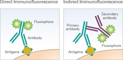 Immunofluorescence Assays Principle Ibidi From ibidi.com
Fix the cells with 4 formaldehyde diluted in 1X PBS prepare fresh for 10 min at room temperature fixation time can be. Please also review the datasheet of the antibody and publications using the same antibody for reference. Grow cultured cells on sterile glass cover slips or slides overnight at 37 º C. 1950 and it is a combination of immunofluorescence technique and morphological technology to develop immune fluorescent cells or tissue. Wash the cells by PBS 2 times for 5 min each. Optimizing Immunofluorescence Staining Protocols for Image Quality Immunostaining with Fluorescent Antibodies Uses for immunofluorescence IFwhere an antibody is conjugated to a molecule that fluoresces when excited by lasers include protein localization confirmation of post-translational modification or activation and proximity tocomplexing with other proteins.
Immunofluorescence Staining Protocol from IHC world 1.
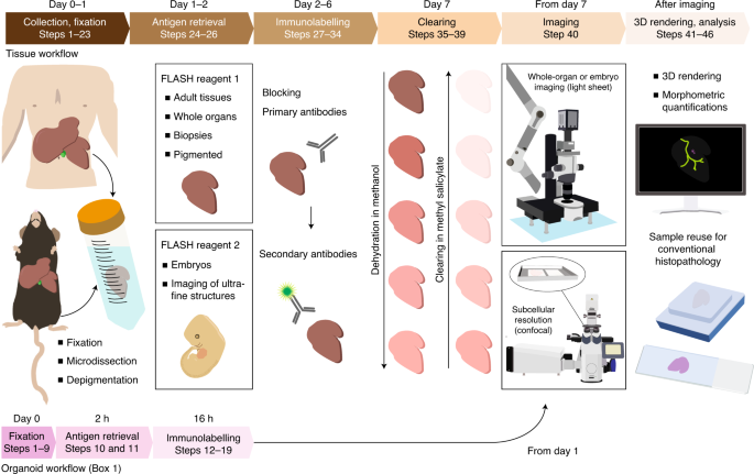
Immunofluorescence stained samples are examined under a fluorescence microscope or confocal microscope. Rinse coverslips well with sterile H 2 O three times 1 h each. Wash briefly with PBS. Antibody molecules for a specific target molecule are exposed to the cell or tissue being investigated. Preparation of Slides. Coat coverslips with polyethylineimine or poly-L-lysine for 1 h at room temperature.
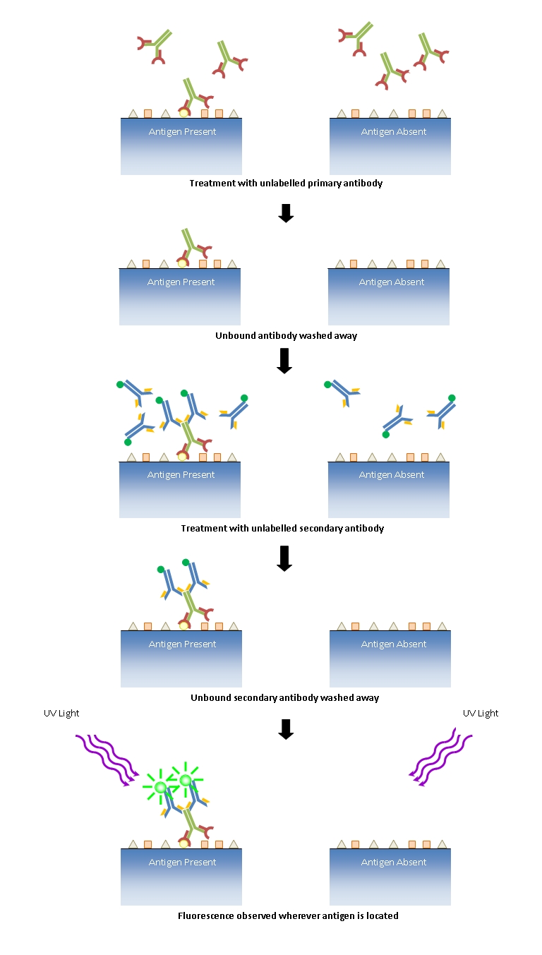 Source: di.uq.edu.au
The following immunohistochemistry IHC protocol has been developed and optimized by RD Systems IHCICC laboratory for fluorescent IHC experiments using frozen tissue samples. Wash briefly with PBS. Optimization of concentration or incubation condition of the primary antibody and the secondary antibody for your own specimen is necessary. Fix the cells with freshly made fixative for 30 min 3. Immunofluorescence staining is a very sensitive method that might require troubleshooting.
 Source: leica-microsystems.com
Fix the cells with freshly made fixative for 30 min 3. Immunocytochemistry and immunofluorescence protocol Abcam. Fix the cells with 4 formaldehyde diluted in 1X PBS prepare fresh for 10 min at room temperature fixation time can be. Stain nuclear with DAPI 05 µgml or Hoechst 01-12 µgml in PBS for 5-10 min. Immunofluorescence staining can be performed on cells fixed on slides and tissue sections.
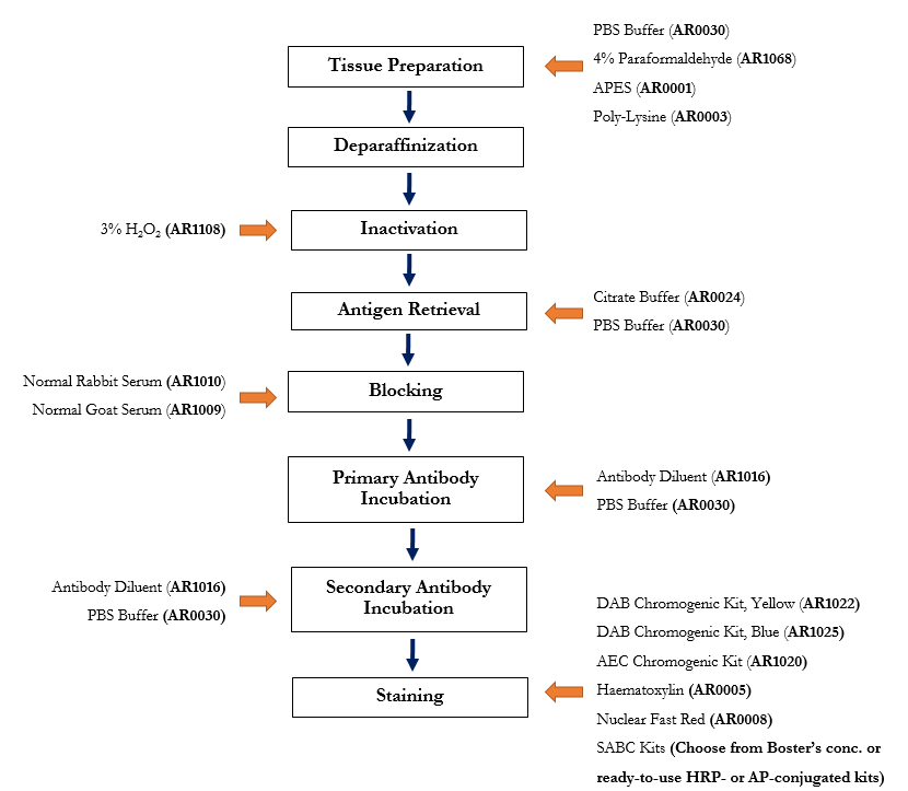 Source: bosterbio.com
Rinse coverslips well with sterile H 2 O three times 1 h each. Coat coverslips with polyethylineimine or poly-L-lysine for 1 h at room temperature. Use suction to remove reagents after each step but avoid drying of specimens between steps. Rinse coverslips well with sterile H 2 O three times 1 h each. Wash the cells by PBS 2 times for 5 min each.
 Source: sciencedirect.com
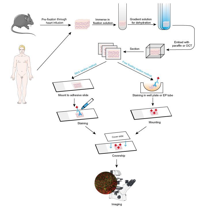
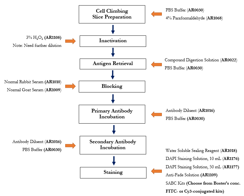
This IHC protocol provides a basic guide for the fixation cryostat sectioning and staining of frozen tissue samples. Immunofluorescence staining can be performed on cells fixed on slides and tissue sections. Rinse the cells with cPBS 2. This is our basic protocol for staining adherent cells in dishes or cells grown on coverslips. Every immunofluorescence staining protocol consists of four major steps cultivation fixation staining imaging which can be subdivided as follows.
 Source: leica-microsystems.com
Place the sterile cover slips in 12 or 24 well plates. Place the sterile cover slips in 12 or 24 well plates. Immunofluorescence Staining Protocol from IHC world 1. Wash briefly with PBS. Immunofluorescence Staining of Cells for Microscopy There are many variations on IF protocols and steps may need to be optimized for different targets or applications.
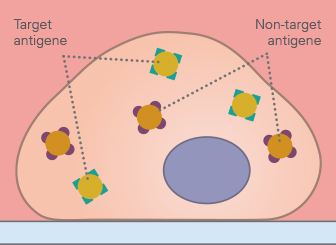 Source: ibidi.com
Immunofluorescence Protocol IF Protocol Immunofluorescence protocol IF protocol Immunofluorescence is one of the widely used techniques in modern biology and medicine and it is developed by Coons et al. Optimizing Immunofluorescence Staining Protocols for Image Quality Immunostaining with Fluorescent Antibodies Uses for immunofluorescence IFwhere an antibody is conjugated to a molecule that fluoresces when excited by lasers include protein localization confirmation of post-translational modification or activation and proximity tocomplexing with other proteins. Protocol Day 1. 10 minutes with 10 formalin in PBS keep wet 5 minutes with ice cold methanol allow to air dry. Guideline procedure for immunofluorescence staining of cell cultures including fixation permeabilization blocking counter-staining and specimen mounting.
 Source: vectorlabs.com
Immunofluorescence staining is a very sensitive method that might require troubleshooting. This is our basic protocol for staining adherent cells in dishes or cells grown on coverslips. 1950 and it is a combination of immunofluorescence technique and morphological technology to develop immune fluorescent cells or tissue. Because fluorescent dyes such as fluorescein and rhodamine can be coupled to antibodies without destroying their specificity the conjugates can complex with antigen and be visualized via fluorescence micro. ICC and IF protocol Preparing the slide 1.
 Source: ultivue.com
Guideline procedure for immunofluorescence staining of cell cultures including fixation permeabilization blocking counter-staining and specimen mounting. Immunofluorescence Staining Protocol. This IHC protocol provides a basic guide for the fixation cryostat sectioning and staining of frozen tissue samples. Fix the cells with 4 formaldehyde diluted in 1X PBS prepare fresh for 10 min at room temperature fixation time can be. Immunofluorescence Staining Protocol from IHC world 1.
 Source: sinobiological.com
Rinse coverslips well with sterile H 2 O three times 1 h each. Place the sterile cover slips in 12 or 24 well plates. Immunofluorescence stained samples are examined under a fluorescence microscope or confocal microscope. Use suction to remove reagents after each step but avoid drying of specimens between steps. Rinse coverslips well with sterile H 2 O three times 1 h each.
 Source: biotium.com
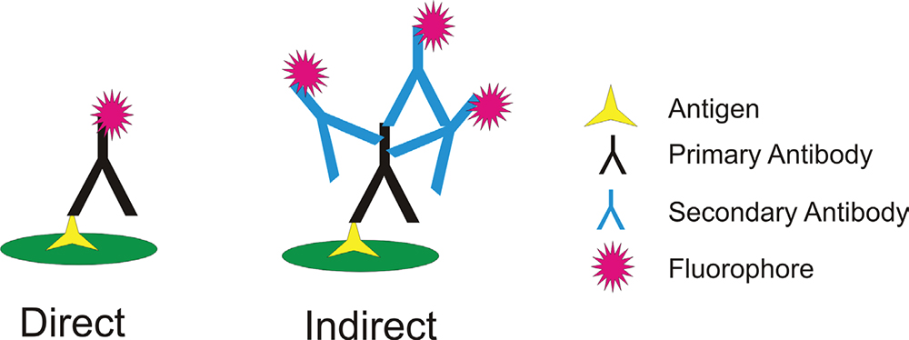
This IHC protocol provides a basic guide for the fixation cryostat sectioning and staining of frozen tissue samples. Immunofluorescence Staining Protocol from IHC world 1. For direct immunofluorescence staining of cells or tissue sections we recommend the use of SCBTs monoclonal antibodies conjugated to fluorescein phycoerythrin Alexa Fluor 488 and Alexa Fluor 647. Protocol Day 1. Plate the cells on the cover slips at a density of 10000 cm2 Day 2 1.
 Source: sinobiological.com
Cell Lines Grow cultured cells on sterile glass cover slips or slides overnight at 37 º C Wash briefly with PBS Fix as desired. Immunofluorescence Staining Protocol. Wash briefly with PBS. Some epitopes may require specific fixation conditions for detection. Protocol for immunofluorescence staining of adhesion cells This is provided as a general protocol.
 Source: leica-microsystems.com
Antibody molecules for a specific target molecule are exposed to the cell or tissue being investigated. Immunofluorescence staining is a very sensitive method that might require troubleshooting. Stain nuclear with DAPI 05 µgml or Hoechst 01-12 µgml in PBS for 5-10 min. Antibody molecules for a specific target molecule are exposed to the cell or tissue being investigated. The following immunohistochemistry IHC protocol has been developed and optimized by RD Systems IHCICC laboratory for fluorescent IHC experiments using frozen tissue samples.
 Source: nature.com
Place the sterile cover slips in 12 or 24 well plates. Guideline procedure for immunofluorescence staining of cell cultures including fixation permeabilization blocking counter-staining and specimen mounting. Immunofluorescence Staining of Cells for Microscopy There are many variations on IF protocols and steps may need to be optimized for different targets or applications. Every immunofluorescence staining protocol consists of four major steps cultivation fixation staining imaging which can be subdivided as follows. Slight changes in the protocol can lead to different results that are no longer comparable.
 Source: sciencedirect.com
This is our basic protocol for staining adherent cells in dishes or cells grown on coverslips. Immunofluorescence Protocol IF Protocol Immunofluorescence protocol IF protocol Immunofluorescence is one of the widely used techniques in modern biology and medicine and it is developed by Coons et al. Some epitopes may require specific fixation conditions for detection. Protocol Day 1. Antibody molecules for a specific target molecule are exposed to the cell or tissue being investigated.
 Source: creative-diagnostics.com
This unit provides a protocol for indirect immunofluorescence which is a method that provides information about the locations of specific molecules and the structure of the cell. Rinse coverslips well with sterile H 2 O three times 1 h each. Coat coverslips with polyethylineimine or poly-L-lysine for 1 h at room temperature. Optimization of concentration or incubation condition of the primary antibody and the secondary antibody for your own specimen is necessary. Immunofluorescence Staining Protocol from IHC world 1.
 Source: epigentek.com
Immunofluorescence Staining of Cells for Microscopy There are many variations on IF protocols and steps may need to be optimized for different targets or applications. Grow cultured cells on sterile glass cover slips or slides overnight at 37 º C. Guideline procedure for immunofluorescence staining of cell cultures including fixation permeabilization blocking counter-staining and specimen mounting. This is our basic protocol for staining adherent cells in dishes or cells grown on coverslips. Preparation of Slides A.
 Source: bosterbio.com
Grow cultured cells on sterile glass cover slips or slides overnight at 37 º C. Immunofluorescence staining can be performed on cells fixed on slides and tissue sections. Preparation of Slides A. 10 minutes with 10 formalin in PBS keep wet 5 minutes with ice cold methanol allow to air dry. Cell Lines Grow cultured cells on sterile glass cover slips or slides overnight at 37 º C Wash briefly with PBS Fix as desired.
 Source: ibidi.com
Immunofluorescence staining is a very sensitive method that might require troubleshooting. Because fluorescent dyes such as fluorescein and rhodamine can be coupled to antibodies without destroying their specificity the conjugates can complex with antigen and be visualized via fluorescence micro. 1950 and it is a combination of immunofluorescence technique and morphological technology to develop immune fluorescent cells or tissue. Immunofluorescence Protocol IF Protocol Immunofluorescence protocol IF protocol Immunofluorescence is one of the widely used techniques in modern biology and medicine and it is developed by Coons et al. Discard the cell culture medium by inverting the slide and gently tapping it on a paper towel to remove the remaining medium.
This site is an open community for users to share their favorite wallpapers on the internet, all images or pictures in this website are for personal wallpaper use only, it is stricly prohibited to use this wallpaper for commercial purposes, if you are the author and find this image is shared without your permission, please kindly raise a DMCA report to Us.
If you find this site value, please support us by sharing this posts to your preference social media accounts like Facebook, Instagram and so on or you can also bookmark this blog page with the title immunofluorescence staining protocol by using Ctrl + D for devices a laptop with a Windows operating system or Command + D for laptops with an Apple operating system. If you use a smartphone, you can also use the drawer menu of the browser you are using. Whether it’s a Windows, Mac, iOS or Android operating system, you will still be able to bookmark this website.